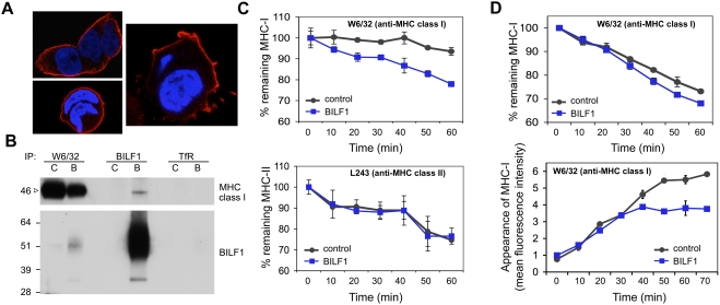
Figure 7. BILF1 associates with MHC class I molecules at the cell surface and increases their rate of internalization.
(A) BILF1 is predominantly localized at the cell surface. 293 cells stably transduced with BILF1 retrovirus were grown on glass slides, fixed and permeabilized, then stained with rat anti-HA (3F10) primary antibodies and Alexa Fluor® 594 goat anti-rat IgG. The nuclei were counterstained with DAPI. The stained slides were analyzed with a laser scanning confocal microscope, and the three photographs show different 1 micron-thick sections through representative cells. BILF1 stained red, and the nuclei stained blue. (B) BILF1 and MHC class I molecules co-precipitate at the cell surface. 293 cells (2×10⁶) stably transduced with control (c) or BILF1 (b) retrovirus were incubated with saturating concentrations of antibodies specific for MHC class I (W6/32), TfR (H68.4) or HA tagged BILF1 (3F10) on ice. After washing away excess antibody, the cells were lysed with NP40 detergent buffer, then precipitated with protein A/G beads and subjected to western-blotting as in Fig. 6B, using antibodies specific for MHC class I (HC10) and HA tagged BILF1 (3F10). (C) BILF1 increases the rate of internalization of MHC class I, but not class II, from the cell surface. MJS cells stably transduced with control or BILF1 retrovirus were incubated at 0°C with saturating concentrations of mAb to MHC class I (W6/32; top graph) or MHC class II (L234; bottom graph), then washed and incubated at 37°C for different periods of time. The cells were subsequently stained with PE-conjugated goat anti-mouse IgG antibody, and analyzed by flow cytometry. The mean fluorescence intensities of staining were averaged for triplicate samples, and normalized to the initial time 0 min samples. (D) BILF1 increases the rate of internalization, but not the rate of appearance, of MHC class I at the cell surface. Top graph: 293 cells stably transduced with control or BILF1 retrovirus were incubated at 0°C with saturating concentrations of mAb to MHC class I (W6/32), then treated exactly as for the internalization assay performed with MJS cells in panel C. Bottom graph: replicate aliquots of the saturated W6/32-bound cells were harvested at the indicated time points, and the appearance of new MHC class I molecules was assayed by staining with PE-conjugated W6/32 antibody. The mean fluorescence intensities of staining were averaged for triplicate samples.

