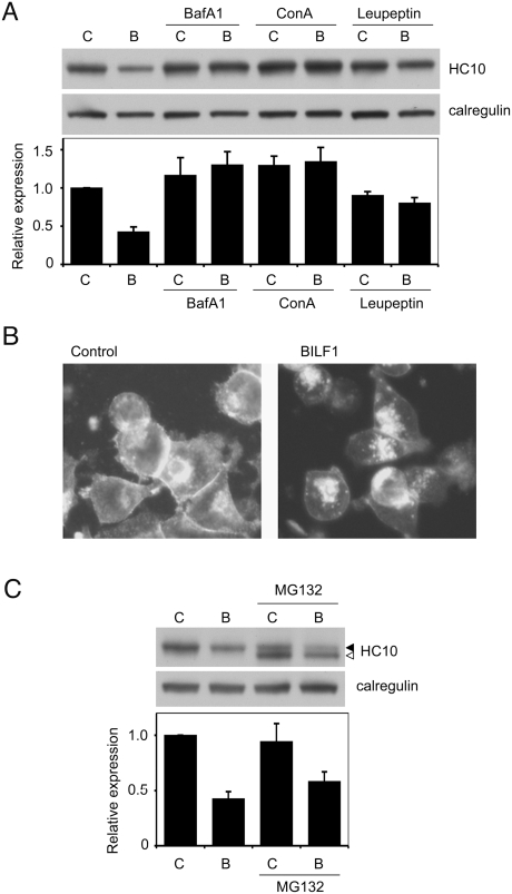
Figure 8. Lysosomal inhibitors block BILF1-enhanced degradation of MHC class I.
(A) 293 cells stably transduced with control- (c) or BILF1- (b) retrovirus were treated with or without Bafilomycin A1, concanamycin A, or leupeptin for 20 hr. Lysates from 2×10⁵ cell equivalents were separated by SDS/PAGE gel, and analyzed by western blotting using antibodies specific for MHC class I (HC10) and calregulin. The blot is one representative of three independent experiments. The histogram shows the mean results (±S.D.) of quantification by densitometry of all the blots from 3 independent experiments, where the densities of the HC10 bands were normalized relative to their own calregulin loading control. (B) 293 cells stably transduced with control or BILF1 retrovirus were treated with concanamycin A (50 nM) for 6 hr prior to fixation and permeabilization with methanol/acetone, then stained with W6/32 primary antibodies and Alexa Fluor® 488 goat anti-mouse IgG secondary antibodies. The photographs were obtained with a conventional fluorescence microscope. (C) 293 cells stably transduced with control (c) or BILF1 (b) retrovirus were treated with or without the proteasome inhibitor, MG132, for 20 hr, and analyzed by western blot as in panel A. The additional, lower molecular weight species detected is probably deglycosylated and/or partially degraded free heavy chain that is normally targeted for proteasomal degradation.

