
Figure 3.
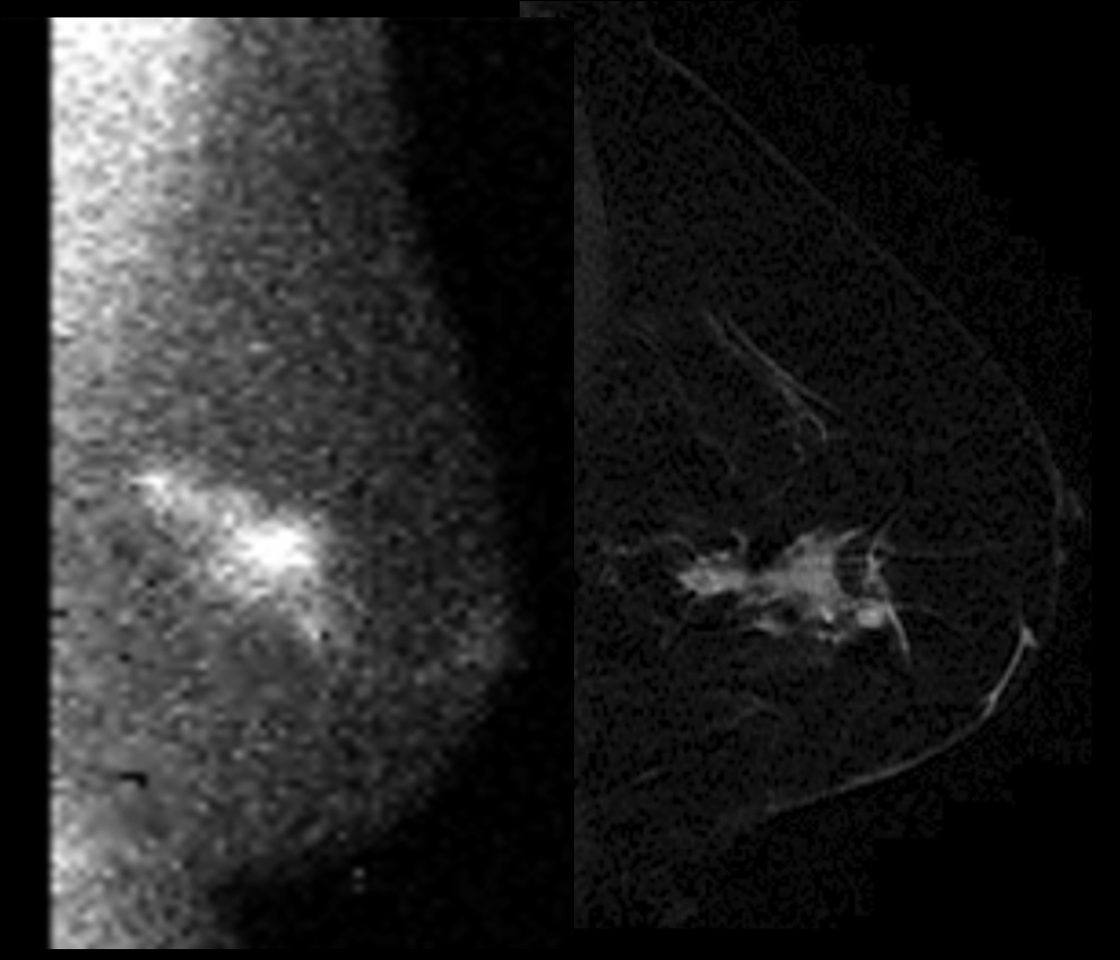
An example of a patient with concordant MBI (left) and breast MRI (Right) findings. MBI shows linear focal abnormal tracer uptake in the posterolateral left breast with orientation towards the nipple. There are 3 foci of intense tracer uptake suggesting a combination of invasive breast cancer and DCIS. MRI shows an irregular enhancing mass in the outer breast measuring 4.6cm × 1.8cm × 2.2cm.