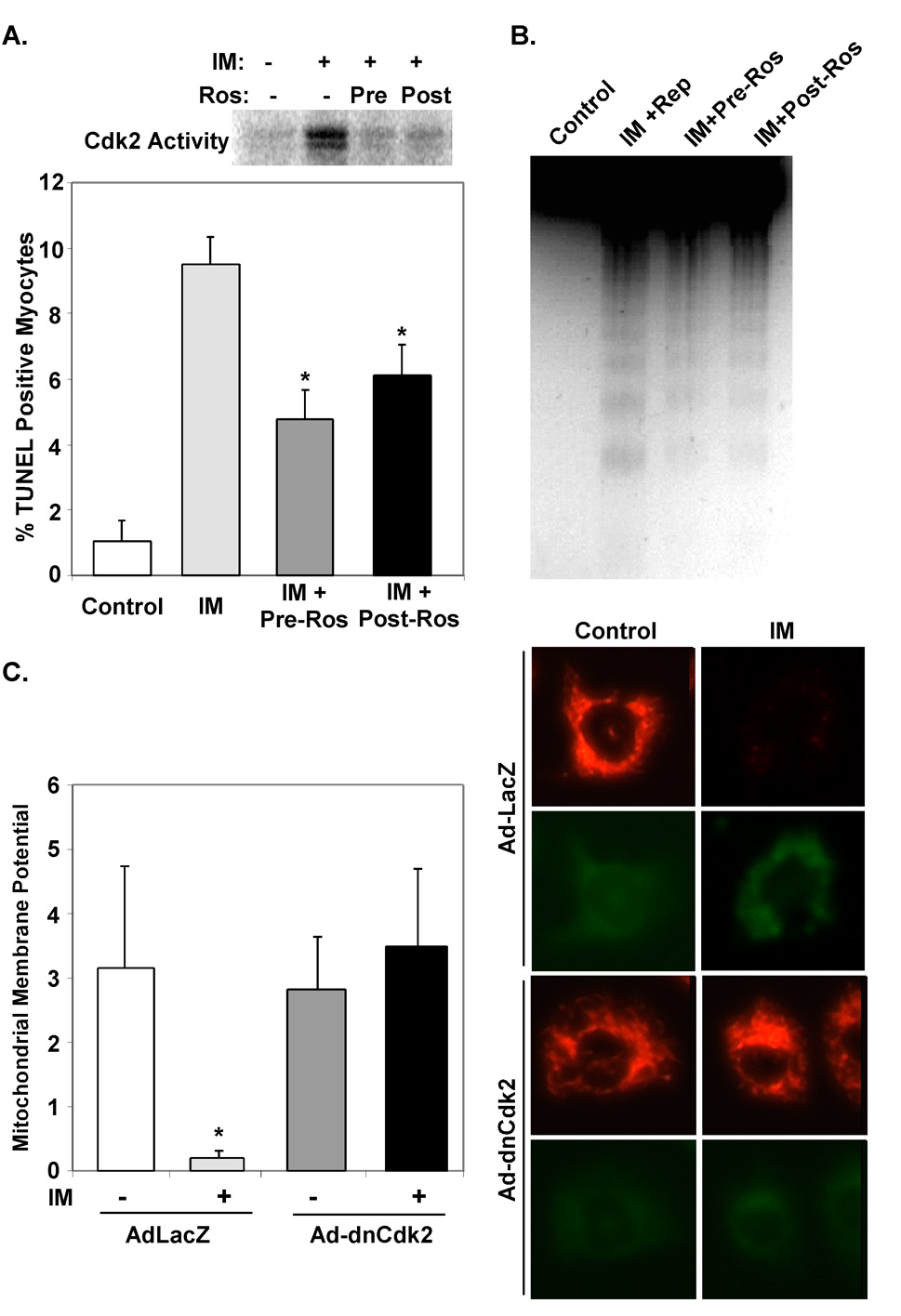
Figure 2. Inhibition of Cdk2 activity attenuates apoptosis and mitochondrial membrane permeability transition in cardiac myocytes.
A & B. Primary cultures of NRVMs were exposed to ischemia media (IM) for 45 minutes and then returned to standard serum free media for the indicated reperfusion times. Roscovitine (10 µM) was added 15 minutes before (Pre) or immediately after (Post) exposure to ischemia medium. Cells were cultured an additional 2 hrs in serum-free media and then protein lysates were prepared. Results of Cdk2 kinase assays are shown (A). TUNEL positive nuclei (A) and DNA laddering (B) was reduced in NRVMs treated with Roscovitine (*P<0.001, n=3 reps). C. NRVMs were isolated and infected with the indicated virus and cultured in serum free media for 48 hours. NRVMs were labeled with mitochondrial dye, JC-1, and exposed to ischemia media (IM) for 45 minutes. Dncdk2 rescued ΔΨm in myocytes subjected to IM when compared to control virus infected myocytes (*P<0.05, n=3 reps).

