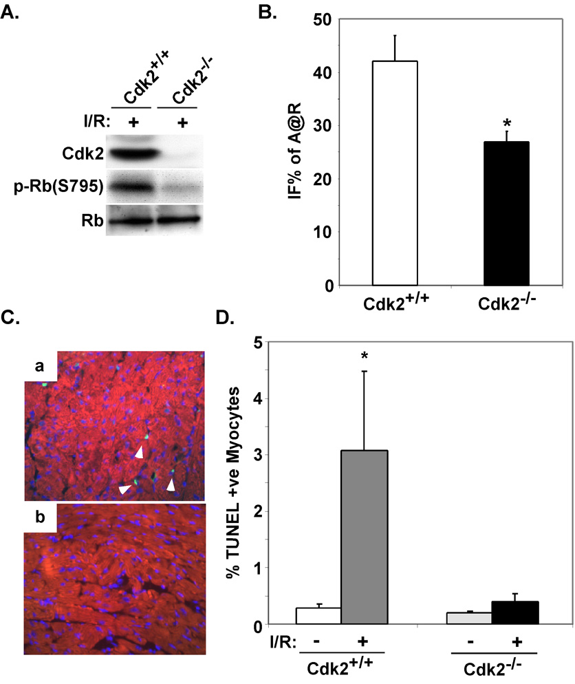
Figure 4. Cdk2-null mice demonstrate reduced infarct size in vivo and apoptosis.
A. Western blots were performed on protein lysates extracted from ventricular tissue from wildtype or Cdk2-null mice subjected to I/R injury. B. Wildtype or Cdk2-null mice were subjected to I/R injury. Mean IFS sizes after 24 hours of reperfusion are shown. (*P<0.05 for Cdk2+/+ versus Cdk2−/− IFS; n=5 per group). C. Immunofluorescent staining for TUNEL (green) and cardiac-specific marker MF20 (red) was performed on myocardial sections from wildtype or Cdk2-null mice subjected to I/R. D. The percentage of TUNEL positive nuclei was quantified on myocardial sections from the indicated genotypes and treatments. The results from examination of at least 3,500 nuclei per animal are shown. (*P<0.05 Cdk2+/+ after I/R versus Cdk2+/+ or Cdk2−/− at baseline and P= 0.05 for Cdk2+/+ versus Cdk2−/− after I/R; n=4 per group).

