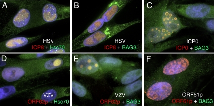
Fig. 1.
Localization of Hsc70 and BAG3 during VZV and HSV infection or ICP0 transformation. MeWo cells were infected with HSV (A and B) or cell-free VZV (D and E) or transformed with a plasmid expressing HSV-ICP0 (C) or VZV-ORF61p (F). Cells were fixed at 6, 24, or 48 hpi, respectively, and the indicated proteins were visualized by indirect immunofluorescence microscopy. Images were captured with a 100× objective and analyzed by volume deconvolution. The individual separated channels are presented as Fig. S1.

