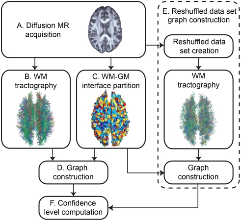
Figure 1. Overview of the whole process.
Overview of the whole process. (A) Acquisition of the diffusion MR images. (B) Tractography in the brain WM. (C) Partitioning of the WM-GM interface into small regions of interest (ROIs). (D) Creation of the original brain connectivity graph using the results of steps B and C. (E) Construction of randomized versions of the original brain connectivity graph (the same partition into ROIs is used). (F) Computation of the confidence level of every edge in the original brain connectivity graph.

