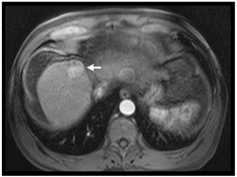
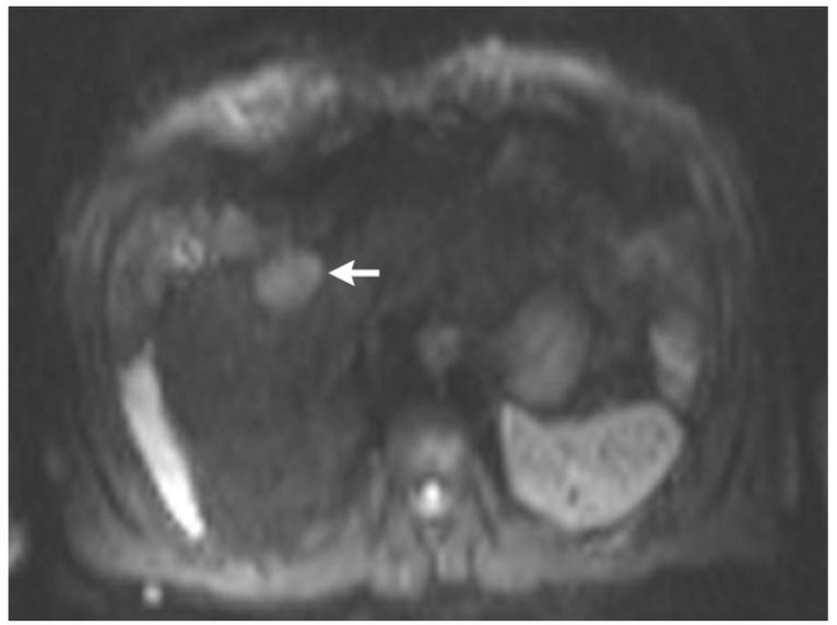
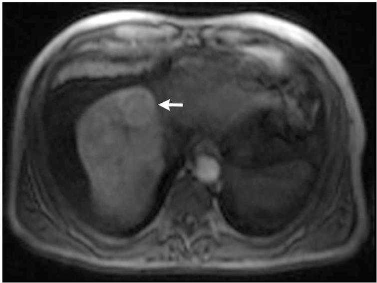
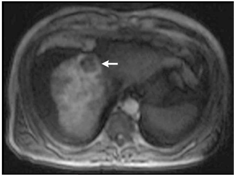
Figure 2.
Post-contrast T1-weighted (a) and diffusion weighted (b) MR scans obtained prior to TACE reveal segment 8 HCC, as shown in figure 1. Pre- (c) and post-TACE (d) 4D TRIP-MRI shows dramatic reduction in tumor perfusion (arrow) after embolization.


