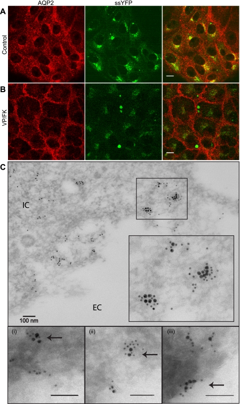
Fig. 2.
Colocalization of ssYFP and aquaporin-2 (AQP2) in LLC-PK1 cells. A: partial colocalization of AQP2 (red) with ssYFP (green) in a perinuclear compartment. B: localization of AQP2 and ssYFP after 15 min of stimulation with vasopressin-forskolin (VP/FK). AQP2 is relocated to the plasma membrane, whereas some ssYFP remains inside the cell, especially in a perinuclear compartment. Images represent native ssYFP fluorescence and immunostained AQP2. Scale bar, 10 μm. C: immunoelectron-microscopic colocalization of AQP2 (15-nm gold) and ssYFP (10-nm gold), as indicated by close association of AQP2 and ssYFP in some intracellular clusters in some regions. Bottom: clusters of colocalized AQP2 and ssYFP (arrows) at higher magnification approaching the membrane (i), at the membrane (ii), and after fusion (iii). IC, intracellular; EC extracellular.

