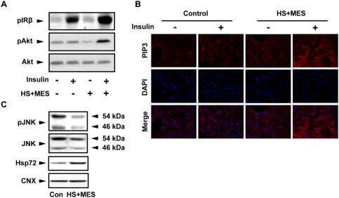
Figure 4. HS+MES improved insulin signaling in the liver of high fat-fed mice.
(A) Liver lysates were extracted 15 weeks after initiation of HS+MES treatment from high fat-fed control and HS+MES-treated mice with or without 5 units of insulin stimulation through inferior vena cava, and were analyzed by Western blotting. (B) Liver tissues were isolated at the 15th week after initiation of treatment from high fat-fed control or HS+MES-treated mice with or without 5 units of insulin stimulation through inferior vena cava. Tissues were dissected in frozen sections, stained with PIP3 and DAPI, and visualized by fluorescent microscope. Scale bars, 100 µm. (C) Liver lysates extracted from control and HS+MES-treated mice were subjected to Western blotting analysis using the indicated antibodies.

