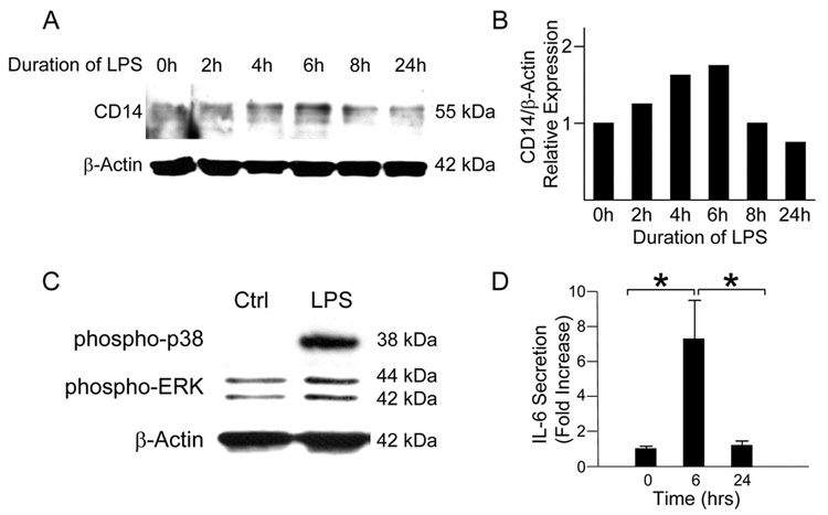
Figure 1. CD14 expression exhibits a transient response to LPS stimulation in IEC-6 cells.
(A) IEC-6 cells were stimulated with LPS (50 µg/ml) for 0 to 24 hours. SDS-PAGE and band density analysis of lysates demonstrates a peak of CD14 protein at 6 hours after treatment followed by a gradual return to near baseline levels by 24 hours. (B) IEC-6 cells stimulated with LPS demonstrate an increase in phosphorylation of the MAPK p38 and ERK after 20 minutes of stimulation. (C) Analysis of the media of LPS-stimulated IEC-6 cells by ELISA demonstrates a significant increase in IL-6 levels at 6 hours after stimulation (1.0 +/− 0.1 vs. 7.1 +/− 2.6 fold increase, *p<0.05). IL-6 levels return to baseline by 24 hours (7.1 +/− 2.6 vs. 1.2 +/− 0.2 fold increase, *p<0.05).

