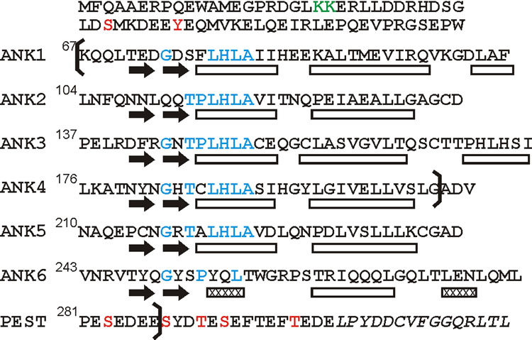
Figure 1.
Amino acid sequence of human IκBα showing the positions of the ankyrin repeats and the PEST domain. Positions of secondary structure elements in the X-ray crystal structure of IκBα in complex with p65(19-321)/p50(248-350)10 are indicated below the sequence. The positions of the hairpin structures in each repeat are indicated by black arrows, and the helices by rectangles, with 310 helices shown hatched. Residues that correspond to the ankyrin repeat consensus are shown in blue. Phosphorylation sites are indicated in red, and a ubiquitination site in green. The boundaries of the constructs used in this study (67-287, 67-206) are shown by brackets. The C-terminal sequence thought to be involved in ubiquitin-independent proteasome degradation53 is shown in italics.

