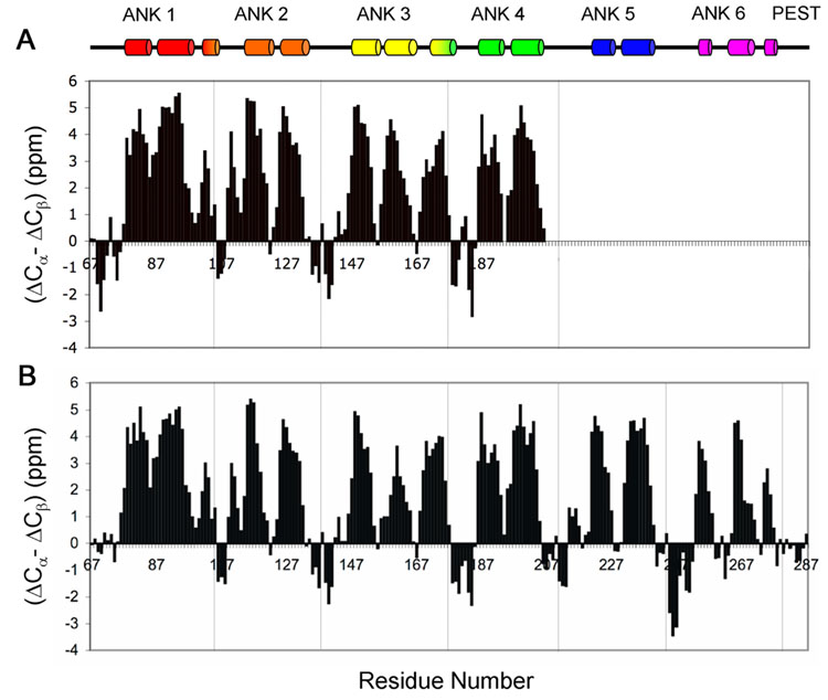
Figure 6.
Secondary structure, evaluated by the parameter (ΔCα - ΔCβ) plotted versus residue number for A. IκBα(67-206). The chemical shift values of 13Cα and 13Cβ were obtained for free [13C, 15N]-labeled IκBα(67-206). B. IκBα(67-287) in complex with p50(248-350)/p65(190-321). The chemical shift values of 13Cα and 13Cβ were obtained for the complex of [2H, 13C, 15N]-labeled IκBα(67-287) with highly deuterated p50(248-350)/p65(190-321). ΔCα and ΔCβ were calculated from the difference between the experimental 13Cα and 13Cβ chemical shifts and the corresponding random coil values. The value of (ΔCα - ΔCβ) for each residue represents the average of three consecutive residues, centered at the particular residue32. The α-helices determined by X-ray for the complex are indicated at the top for comparison.

