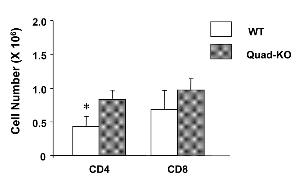
Figure 4. Donor T cell expansion in vivo.
On day 7 after BMT, spleen cells harvested from the recipients received wild type or Quad-KO T cells, and staining with mAbs against donor-maker (H-2Dd) and CD4 or CD8. The cells were analyzed by FACSVantage SE and the numbers of donor-derived CD4 or CD8 in the host spleen (n = 3–4 mice/ group) were calculated. The difference of the number of donor CD4+ cells was statistically significant. Data shows mean ± SEM. * P<0.05.

