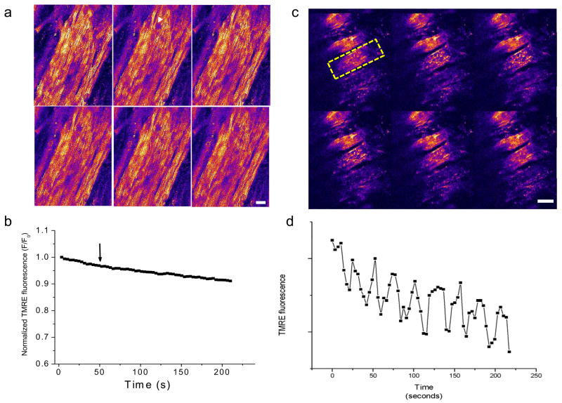
Figure 1.
Mitochondrial membrane potential oscillations in myocytes of the intact heart perfused with Ca2+-free buffer. a, b) The ΔΨm signal (TMRE fluorescence) was stable over many minutes in hearts perfused with modified Tyrode’s containing 0.5 mM Ca2+ even after a localized laser flash was applied (white arrowhead in panel a, arrow in panel b). c, d) After 2–3 min of normoxic perfusion with Ca2+-free Tyrode’s, ΔΨm became unstable and spontaneous oscillations were observed. Sustained oscillations in ΔΨm were observed for several minutes in the cell indicated by the yellow dashed line in panel c. Both intracellular and intercellular heterogeneity of ΔΨm is evident in the epicardial optical sections. Scale bars in a and b equal 20 microns.

