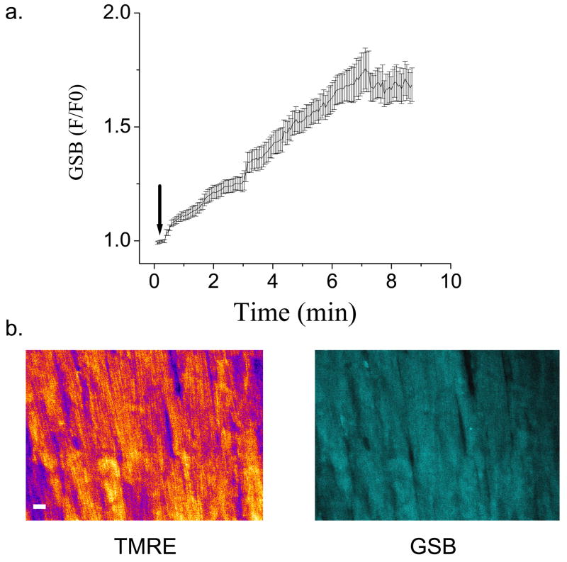
Figure 5.
Measurement of reduced glutathione in the intact heart using monochlorobimane. Intact guinea pig hearts loaded with TMRE were perfused at a constant flow with oxygenated Tyrode’s solution for a 20-min stabilization period (to obtain baseline NADH fluorescence signal). Subsequently, 50μM MCB was added (at arrow in panel a) in a total perfusate volume of 10ml and recirculated until a steady state fluorescence was obtained, at which point, MCB-free Tyrode’s perfusion was restored. Intracellular uptake of MCB was observed as an increase in the 480nm fluorescence emission above the NADH baseline. a) The kinetics of GSB production in the intact heart during MCB loading. b) The intracellular distribution of TMRE and GSB in images of the myocardial syncytium. Epicardial images (about 100μm depth) were acquired at 3.5-second intervals. Scale bar in b equals 20 microns.

