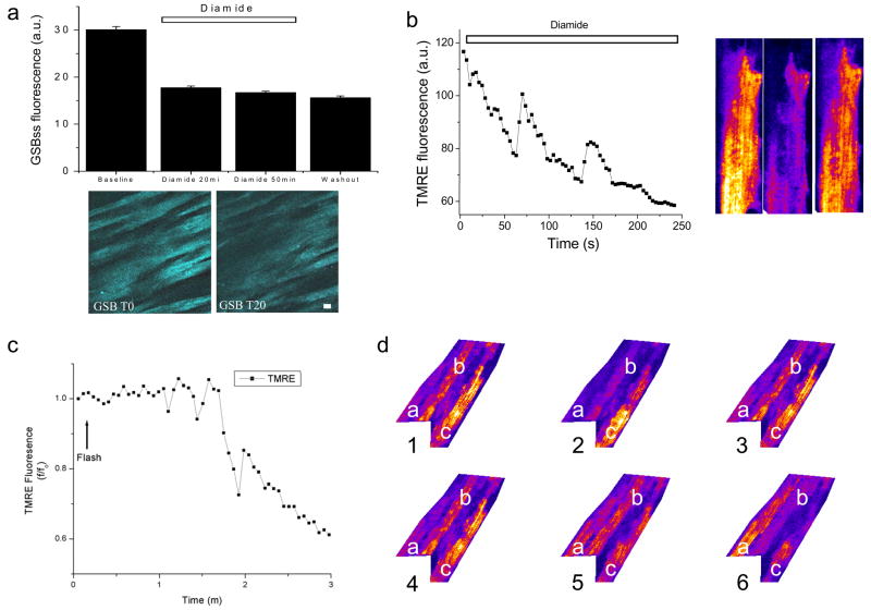
Figure 6.
Depletion of reduced glutathione elicits mitochondrial ΔΨm oscillation in the intact heart. During normoxic perfusion, 1mM diamide, a thiol pro-oxidant, and 1mM BSO, an inhibitor of glutathione synthesis, were added to the perfusate to deplete the intracellular GSH pool. a) The average steady-state GSB fluorescence (GSBss) decreased in the presence of diamide and BSO when whole microscopic fields (below) were analyzed. The decreased GSBss levels persisted after washout (W) of the diamide and BSO. After whole-heart treatment with diamide and BSO, oscillations in ΔΨm were observed either spontaneously (b) or after a laser flash (c). d) A series of sequential images (1–6) of 3 representative cells in one optical field of the intact heart. Cell “a” is initially depolarized and repolarizes by image 6. Directly adjacent to cell “a”, cell “b” undergoes two ΔΨm oscillations over the same time period. Cell “c” is polarized in image 1, but partially depolarizes by the end of the sequence.

