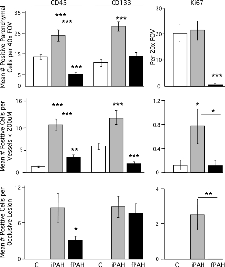
Fig. 1.
Quantification of CD45-, CD133-, and Ki67-positive cells in human pulmonary arterial hypertension (PAH) lung tissue. CD45, CD133, and Ki67 were localized in human lung tissue by antibody staining. Colocalization with vascular structures was performed by costaining with α-smooth muscle actin (α-SMA). 4′,6′-Diamidino-2-phenylindole (DAPI) was used as the nuclear stain. Thirty-five to fifty random fields of view (FOV) were scanned to identify a minimum of 10–12 vessels and occlusive lesions per specimen. Each FOV was counted in a blinded manner using a ×40 or ×20 objective. Data are presented as means ± SE counted by diagnosis. Groups were distinguished as: control (C), white; idiopathic PAH (iPAH), gray; and familial PAH (fPAH), black. *Indicates difference from control P < 0.05 unless indicated with a bar. **P < 0.01; ***P < 0.001.

