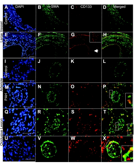
Fig. 3.
CD133-positive progenitor cells increased in PAH tissue. CD133-positive progenitor cells were identified in lung tissue by antibody staining to detect CD133 and vascular structures by α-SMA. DAPI was used as the nuclear stain. CD133 was not detected in association with large control vessels (A–D), arterioles (I–L), or parenchymal tissue. In contrast, large vessels from PAH patients displayed intimal remodeling and a high number of CD133-positive cells (E–H; G, inset, enlarged intimal area). Arterioles from PAH tissue (M–P) had CD133 localized both adjacent to and infiltrating the smooth muscle layers. High levels of CD133 expression were detected within and adjacent to occlusive lesions (Q–X). CD133 coexpression with α-SMA was detected and indicated by yellow in the merged panels. Scale bars = 75 μm.

