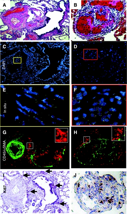
Fig. 6.
Cells present in PAH vascular lesions had normal ploidy. Remodeled arterioles with SMC hypertrophy (A) and intimal occlusive lesions (B) were identified in human lung tissue specimens by H&E stain. Serial sections were then analyzed for ploidy using chromosome probe sets (C–F) and CD45 (red) and α-SMA (green) expression (G and H). Boxes in C and D were enlarged to E and F where fluorescence in situ hybridization probe sets may be visualized in the nuclei. Magnification, ×20. I and J: corresponding Ki67 stain on serial sections. Arrows point to areas of positive Ki67-stained nuclei.

