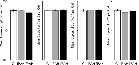
Fig. 7.
Ploidy analysis of vascular lesions in human PAH lung Tissue. Specific arterial and occlusive lesions were identified on the H&E-stained sections that were serial to the sections used for chromosomal analyses. Fluorescence in situ hybridization (FISH) assay was performed with the multicolor, 4 multitarget LAVysion probes. Complete chromosomal analysis was conducted on 10 PAH specimens, 5 iPAH and 5 fPAH, in 4–6 areas per specimen with 10–45 cells analyzed per area. In addition, 5 areas with histologically “normal” lung tissue and vasculature were selected and analyzed as controls for chromosomal gain and loss variability. No significant change in ploidy content from 2 N was identified with any of the 4 probes (y-axis). Data are presented as means ± SE counted by diagnosis. Groups were distinguished as: control, white; iPAH, gray; and fPAH, black.

