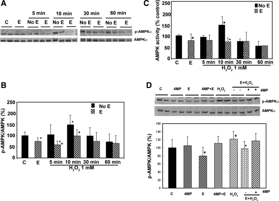
Fig. 3.
Effect of ethanol treatment on the activation of AMPK by oxidative stress. H4IIEC3 cells were grown as described for Fig. 1, followed by treatment with control (C) or ethanol (E) (50 mM); H2O2 1 mM (A) for the indicated duration of treatment, either with pretreatment with ethanol 24 h before H2O2 exposure (E) or without ethanol pretreatment (No E). Western blots were quantified by a PhosphorImager and ImageQuant software analysis (results shown as a bar graph, B). Ethanol treatment for 24 h decreased the p-AMPK/AMP ratio by 25% compared with control (P < 0.05). H2O2 treatment increased the expression of p-AMPK with peak effect occurring at 10 min after treatment (1.5-fold induction over control, P = 0.002). The effect of H2O2 at 5- and 10-min treatment on AMPK phosphorylation was reduced by 43% (P = 0.01) and 33% (P = 0.006), respectively, when the cells were pretreated with ethanol for 24 h. C: AMPK enzyme activity was analyzed with a synthetic peptide substrate, and it correlated well with the level of p-AMPK in B. D: H4IIEC3 cells were treated as indicated with control, 4-methylpyrazole (4MP) (0.1 mM), ethanol (50 mM), ethanol (50 mM) + 4-methylpyrazole (0.1 mM), H2O2 1 mM for 10 min, E + H2O2, pretreatment with ethanol (50 mM) followed by H2O2 1 mM for 10 min, pretreatment with ethanol (50 mM) + 4-methylpyrazole (0.1 mM) for 24 h followed by H2O2 1 mM for 10 min. 4MP did not significantly affect the phosphorylation level of AMPK. However, it abolished the inhibitory effect of ethanol on H2O2-induced AMPK phosphorylation. Means ± SD (the blot is representative of 3 blots from 3 individual experiments) are shown. *Significant difference vs. control; +significant difference compared with pretreatment with ethanol. P < 0.05 by one-way ANOVA.

