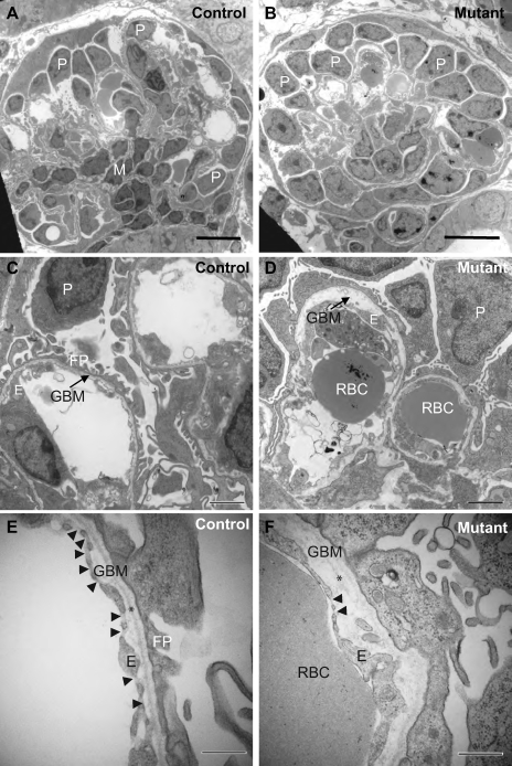
Fig. 4.
Disruption of the filtration barrier before podocyte apoptosis. A, C, and E: electron microscopic micrographs of control glomeruli at P1. B, D, and F: electron microscopic micrographs of mutant glomeruli in similar positions at P1. Well-defined podocyte foot processes were present in the control glomeruli but essentially absent in the mutant glomeruli (C–F). The level of endothelial fenestration was reduced (C–F). Arrowheads in E and F point to endothelial fenestration. Glomerular basement membrane (GBM) thickness varied greatly in mutants. The distance between podocytes and the endothelium was greatly increased (D and F), likely due to the apparent focal GBM splitting (arrow in D). The lamina densa was visible in the control (* in E) but was discontinuous and even absent in many areas (* in F). Scale bars = 10 μm (A and B), 2 μm (C and D), 500 nm (E and F). P, podocyte; E, endothelial cell; M, mesangial cell; RBC, red blood cell; FP, foot processes.

