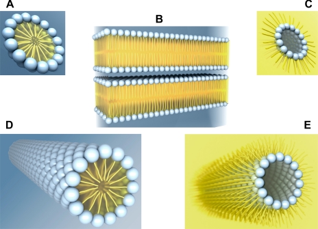
Fig. 2.
Lipid polymorphism. Schematic illustration of a micelle (A), bilayers (B), an inverted micelle (C), and a hexagonal (HI, D) or an inverted hexagonal (HII, E) cylinder. Each phospholipid molecule consists of a hydrophilic (water-loving) head group (blue spheres) facing an aqueous environment (blue background) and 1 or 2 hydrophobic (water-fearing) carbon tails (yellow) facing a lipid environment (yellow background).

