Fig. 1.
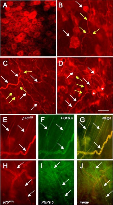
p75NTR and pan-neuronal marker, protein gene product 9.5 (PGP9.5), immunoreactivity (IR) in urinary bladder whole mount preparations with the urothelium/suburothelium dissected from the detrusor smooth muscle. A: confocal image of p75NTR-IR in urothelial cells. B: epifluorescence image of p75NTR-IR suburothelial nerve fibers (yellow arrows) in close proximity to p75NTR-IR urothelial cells (white arrows). C: p75NTR-IR in suburothelial nerve fibers (white arrows) and vasculature (yellow arrows). D: p75NTR-IR in vasculature (yellow arrows), nerve fibers (white arrows), and urothelial cells out of the focal plane (*). p75NTR-immunoreactive suburothelial fibers (arrows E, H) also expressed PGP9.5-IR (arrows F, I). G and J: merged images of E, H and F, I, respectively. Calibration bar represents 100 μm.

