Fig. 2.
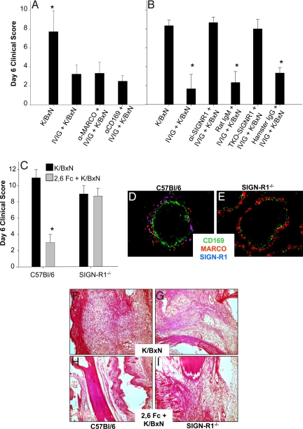
The marginal zone macrophage receptor SIGN-R1 is required for IVIG anti-inflammatory activity. (A) Mice were administered blocking antibodies to marginal zone receptors CD169 (α-CD169), and MARCO (α-MARCO), treated with IVIG followed by K/BxN sera, and footpad swelling monitored. Mean and standard deviation of day 6 clinical scores from 4–5 mice per group are plotted; *, P < 0.05 as determined by ANOVA followed by a Tukey post-hoc test. (B) In a separate experiment, mice were treated with SIGN-R1 blocking antibodies (α-SIGNR1), isotype control antibodies for α-SIGNR1 (Rat IgM), antibodies that transiently knockdown SIGN-R1 expression (TKO-SIGN-R1), or an appropriate isotype control for TKO-SIGN-R1 (Hamster IgG). Next, IVIG and K/BxN were administered, and footpad swelling was monitored over the following week; means and standard deviations of 4–5 mice per group are plotted. *, P < 0.05 as determined by Anova followed by Tukey post-hoc. (C) C57BL/6 and SIGN-R1−/− mice were administered arthritis inducing sera (K/BxN, black bars), some of which received 2,6-Fcs 1 h earlier (2,6-Fc + K/BxN, gray bars). Footpad swelling was monitored over the next seven days in terms of clinical scores. Means and standard deviations of 34 mice per group are plotted. *, P < 0.05 as determined by ANOVA followed by Tukey post-hoc. The splenic marginal zones of C57BL/6 wild type (D) and SIGN-R1−/− (E) mice were examined by immunofluorescence for CD169 (green), MARCO (red), and SIGN-R1 (blue). Monocyte and neutrophil infiltration in the ankle bones of C57BL/6 or SIGN-R1−/− mice were examined in H&E-stained sections 7 days after treatment. Darkly stained nuclei of monocytes and neutrophils infiltrate the ankle bones of both C57BL/6 (F) and SIGN-R1−/− (G) mice treated with K/BxN sera; the inflammatory infiltration is reduced in C57BL/6 mice receiving 2,6-Fc treatment (H) but not in 2,6-Fc-treated SIGN-R1−/− (I) mice.

