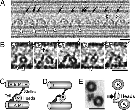
Fig. 2.
Dynein-MT complex in the no-nucleotide state. (A and B) Cryo-positive stain EM images of dynein purified from S. nudus, bound to MTs in the presence of apyrase. The B-MT minus end is at Right. Dynein molecules are regularly arranged in a single layer between 2 MTs. Individual dynein molecules may show a single ring (B, #1–3), a double ring (B, #4), or be unclear (B, #5). Stalks (arrows), the head-A-MT tethers (white arrowheads), and extra densities on the top of the tails (asterisks) are indicated. Some molecules show 2 stalks. (C and D) Interpretation of the single-ring and double-ring images. (E) Cross sections of the dynein-MT complex embedded in Epon812, with our interpretation of the images. The likely viewing direction of our cryo images is indicated. The micrographs in A, B, and Fig. 4 were Gaussian-filtered to reduce noise. (Scale bars: A and B, 20 nm; C, 50 nm.)

