Fig. 3.
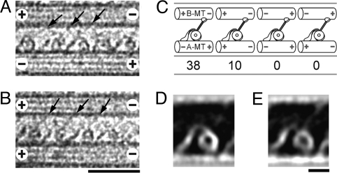
Orientation of dynein relative to the MT polarity. (A and B) Cryo-positive stain EM images of dynein cross-bridging anti-parallel (A) or parallel (B) MTs. (C) Proportion of MT pairs with different polarities. Although the polarity of the A-MT is not uniform, all of the dynein molecules are oriented in the same way with respect to the B-MT, with the heads and stalks to the B-MT minus-end side of the tail. The observed head/tail arrangement agrees with that suggested from the QFDE images of dynein in axonemes (11, 12), but inconsistent with the assignment in a recent report on the Chlamydomonas dynein-MT complex (18). (D and E) Averaged images of dynein cross-bridging 2 anti-parallel MTs (D; n = 30) or parallel MTs (E; n = 31), showing no detectable differences. (Scale bars: A and B, 50 nm; D and E, 10 nm.)

