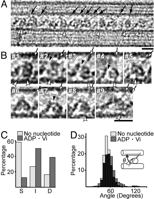
Fig. 4.
Structural changes of dynein with ADP·Vi. (A and B) Cryo-positive stain EM images in the ADP·Vi state, with the B-MT plus end at Left. Individual dynein images (B) show a double ring (#1–6), a single ring (#7), or an intermediate conformation (#8). Most stalks are clearly tilted toward the B-MT minus end (arrows). (C) Populations of dynein molecules in the no-nucleotide and ADP·Vi states showing a superimposed, single ring (S), a double ring (D), or, intermediate or unclear structures (I). (D) Distribution of stalk angles (θ) with respect to MTs. The averaged angles are 54.0 ± 8.7° (n = 451) and 58.6 ± 14.6° (n = 490) for the no-nucleotide and ADP·Vi states, respectively. (Scale bars: 20 nm.)

