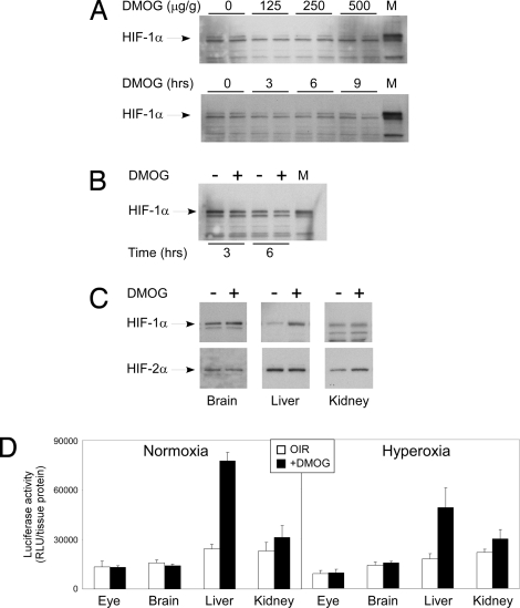
Fig. 6.
Analysis of HIF-1,2 expression. (A) Western blot of ocular HIF-1α shows no dose or time response to DMOG. Western blot of both retina and total eye lysate from the P6 mouse using four different commercial antibodies to HIF-2α showed no trace of this protein in the P6 retina or whole eye lysate (data not shown). The lack of change in HIF-1α stability was also noted in hyperoxia at 3 h and 6 h after DMOG injection, shown in B. On the other hand, (C) hepatic HIF-1α and renal HIF-2α demonstrated increased stability after DMOG injection. To confirm this finding, the luciferase–oxygen degradation fusion protein transgene (luc-ODD) was used to identify luminescence and hence stability of proteins containing the ODD after i.p. DMOG injection. At P6, (D) DMOG injection clearly demonstrates stabilization of hepatic luc-ODD in comparison with eye and brain, matching the Western blot results in A, B, and C. Taken together, the ELISA, Western blot, and luc-ODD analysis strongly correlate to suggest that the P6 mouse liver is a target of i.p. DMOG, and it is the liver that may protect the retinal vasculature once DMOG stimulates hepatic HIF-1α stabilization.

