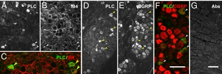
Fig. 5.
Expression of PLCβ3 in human DRG and double-staining experiments as well as absorption control. (A–F) PLCβ3-IR cell bodies are present in human DRGs (A and D) and express IB4 (A–C) or CGRP (D–F). Arrowheads indicate coexistence between PLCβ3 and IB4 (C) or CGRP (F), respectively. (G) After incubation with control serum, no fluorescent neurons can be observed. (Scale bars: 100 μm, A–B–D–E– G; C–F).

