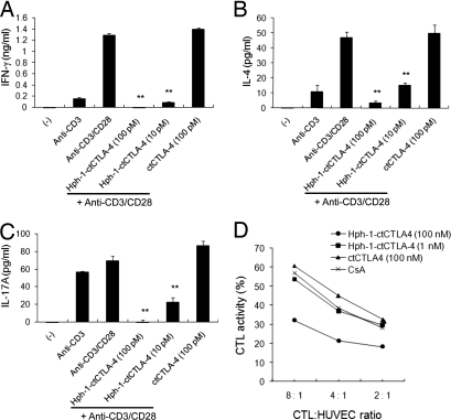
Fig. 1.
Inhibition of T cell activation by Hph-1-ctCTLA-4. After Hph-1-ctCTLA-4 or ctCTLA-4 was incubated with mouse splenocytes for 1 h, cells were stimulated with plate-bound anti-CD3 and soluble anti-CD28 antibodies for 24 h. (A) IFN-γ, (B) IL-4, and (C) IL-17A levels in the culture media were analyzed by ELISA. (D) HUVEC-specific human primary CTL was generated, and Hph-1-ctCTLA-4-transduced CTL was cocultured with calcein-treated HUVEC cells for 4 h. CTL killing activity was calculated as killing percentage. Bars represent mean ± SD of three independent experiments. *P < 0.05, **P < 0.01 compared with the activated T cell group.

