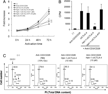
Fig. 3.
Hph-1-ctCTLA-4 suppresses proliferation and cell cycle of activated T cells. (A) After each protein was incubated with Jurkat T cells for 30 min, cells were stimulated with plate-bound anti-CD3 antibody and soluble anti-CD28 antibody for 24–72 h. WST-8 dye was added to each well to stain total live cells. (B) After splenocytes were incubated with each protein and stimulated as described above, proliferation of cells was indicated by the number of thymidine-incorporating cells. (C) After splenocytes were stimulated as described above for 3 days, cells were fixed and intracellular DNA was stained by PI, and the cell cycle was analyzed by FACS. Bars represent mean ± SD of three independent experiments. *, P < 0.05; **, P < 0.01 compared with the activated T cell group.

