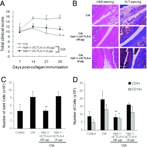
Fig. 6.
Transdermal application of Hph-1-ctCTLA-4 in the collagen-induced arthritis model. (A) Seven days after boost injection of CII antigen, Hph-1-ctCTLA-4 and ointment mixture were administered on the joint skin of the mice five times a week for 4 weeks. Severity of the arthritis symptom was measured by clinical scoring. (B) Each joint sample was analyzed for histological analysis by H&E and M-T staining to examine inflammatory tissue damage and collagen deposition, respectively. (C) The number of total joint cells and (D) the number of CD3+ or CD19+ cells were calculated with the percentage of each staining and the total number of cells by FACS. Bars represent mean ± SEM of two independent experiments from four mice per group. *, P < 0.05; **, P < 0.01 compared with the control CIA group.

