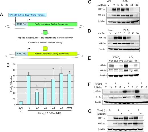
Fig. 1.
Inhibition of HIF-1α and HIF-2α by cardiac glycosides. (A) Hep3B cells were stably co-transfected with: p2.1, a plasmid containing firefly luciferase coding sequences downstream of a hypoxia response element (HRE) and the SV40 early region promoter (Top); and pSV-Renilla, a plasmid containing Renilla luciferase coding sequences downstream of the SV40 early region promoter. The ratio of firefly/Renilla luciferase activity in cells exposed to nonhypoxic (20% O2) or hypoxic (1% O2) culture conditions was determined. (B) The effect of the known HIF-1 inhibitor 17-AAG on the ratio of firefly/Renilla luciferase activity in hypoxic cells was determined; mean ± SD (n = 3) are shown. *, P < 0.05 compared to untreated (Student's t test). (C and D) Hep3B cells were exposed to vehicle (V) or the indicated concentration (nM) of ouabain (Oua) or proscillaridin A (Pro) for 24 h under hypoxic (1% O2) or nonhypoxic (20% O2) conditions and cell lysates were subjected to immunoblot assays for HIF-1α, HIF-2α, and β-actin. (E) PC3 cells were exposed to vehicle (V), 50 nM ouabain (Oua), or 50 nM proscillaridin A (Pro) for 24 h. (F) Hep3B cells were exposed to vehicle (V), 50 nM proscillaridin A (P), or 100 nM ouabain (O) under hypoxic conditions for the indicated time and immunoblot assays were performed. (G) Hep3B cells were exposed to vehicle (−) or 100 nM digoxin (+) under hypoxic conditions for the indicated time and immunoblot assays were performed.

