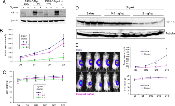
Fig. 6.
Effect of digoxin on HIF-1α expression and tumor xenograft growth. (A) Parental P493-Myc cells and a subclone expressing firefly luciferase (P493-Myc-Luc) were cultured at 20% or 1% O2 for 24 h in the presence of vehicle (−) or 100 nM digoxin (+). (B–D) 2.5 × 10⁷ P493-Myc cells were implanted in s.c. tissue on the flanks of SCID mice (n = 4–5 in each group), which were treated with daily i.p. injections of 0, 0.5, or 2 mg/kg of digoxin in saline, starting 3 days before tumor cell implantation. Tumor volume was determined every 3 days based on caliper measurements; means ± SEM are shown. *, P < 0.05 (Student's t test) (B). Body weight was determined every 5 days; means ± SEM are shown (C). Tumors were harvested on day 23, and tissue lysates were subjected to immunoblot assays (D; each lane represents an independent tumor). (E) P493-Myc-Luc cells were implanted in s.c. tissue on the flanks of SCID mice (n = 5 each), which were treated with daily i.p. injections of saline or digoxin (2 mg/kg) starting 3 days before tumor cell implantation. Luciferase activity was measured on day 8 after tumor cell implantation (Left). Tumor volume (Top Right) and body weight (Bottom Right) measurements were performed on the indicated days; mean ± SEM are shown. [*, P < 0.05 (Student's t test).]

