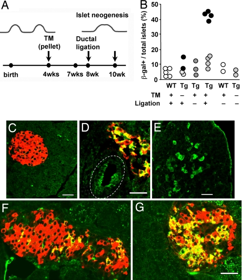
Fig. 4.
Testing hypothesis of progenitors in adult pancreatic regeneration in response to injury. (A) To determine whether new islets are formed in response to injury from duct cells of adult animals, the tamoxifen-inducible CAII-CreERTM double-Tg mice were implanted with 3-week tamoxifen pellets (TM) at 4 weeks of age, had washout period for about 1 week, had duct ligation at 8 weeks, and were killed 2 weeks later. (B) Quantification of positive islets/total islets, with each solid circle representing the mean of a single animal; for ligated Tg, values for pancreas distal to the ligation (dark circles) and for nonligated portion (light circles) are given. The last 2 columns are WT and double Tg at 6 months age, showing the stability of transgene without tamoxifen. In controls or the nonligated portion of the Tg with tamoxifen and ligation (C), there was little β-galactosidase (green) positivity, but distal to ligation in experimentals, some areas had β-galactosidase-marked ducts (D), acini (E), and islets (D, F, and G). In ligated portions, β-galactosidase+ insulin-positive cells were 23.6% ± 2.2% of all insulin-positive cells (mean: 673/2,997 insulin-positive cells; 4 animals), nonligated portions of the same animals were 5.5% ± 2.0% (mean: 110/821 insulin-positive), and in controls were 1.3% ± 1.2% (mean: 11/1,055 cells, 3 WT animals) and 0.9% ± 0.5% (mean: 17/1,785 cells, 4 Tg animals). (D) In this main duct (dashed line), Cre recombination occurred in about half the cells. (E) Expression of β-galactosidase was seen in acinar cells, often localized as to suggest new lobe formation. (F and G) The proportion of positive cells within the marked islets varied as seen in these islets. Shown are expression of β-galactosidase (green), insulin (red), and newly formed beta cells (yellow). (Scale bar, 50 μm.)

