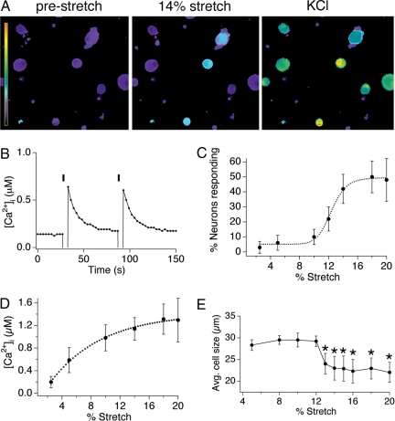
Fig. 1.
Radial stretch activates a subset of trigeminal sensory neurons. (A) Calcium responses of dissociated mouse trigeminal neurons to 14% radial stretch (Middle) and 140 mM KCl ringer's solution (Right). Scale bar indicates the intracellular calcium concentration, ranging from 0.1 to 4 μM [Ca2+]i. (B) Calcium response in a representative cell to two applications of a 14% radial stretch stimulus. (C) Dose–response curve displaying the percentage of neurons activated by varying magnitudes of stretch. (D) Dose–response curve displaying the magnitude of the calcium response triggered by varying magnitudes of stretch. (E) The mean diameter of responding neurons is plotted versus stretch magnitude. For all experiments, n ≥200 cells per point, obtained from a minimum of three different neuronal preparations. All data are reported as means ± SEM.

