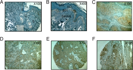
Fig. 6.
Representative immunohistochemistry staining of Srx in normal human skin (A and B), squamous cell carcinoma (C), sweat gland carcinoma (D), basal cell carcinoma (E), and melanoma (F). Note the positive (+) staining of basal cell layer in (A) and (B); (C) and (D), strong positive (++); (E) and (F), very strong positive (+++). BCL, basal cell layer; D, dermis; E, epidermis; GCL, granular cell layer; S, subcutis; SCL, squamous cell layer.

