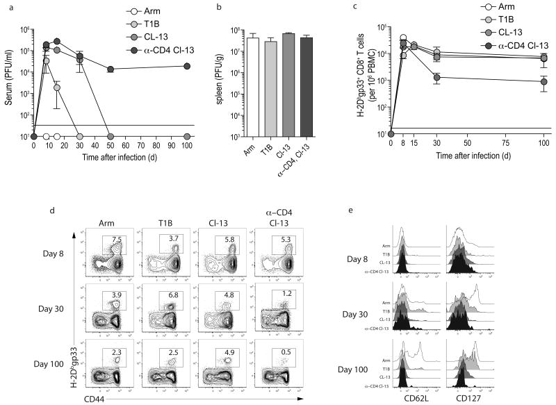
Figure 2. Four types of LCMV infection.
(a) C57BL/6 mice were infected with LCMV Arm, T1B, or clone 13. Also a group of mice was depleted of CD4 T cells with an injection of 200 μg of anti-CD4 (GK1.5) one day prior and one day after infection with LCMV clone 13 (anti-CD4 plus clone 13). Serum was collected from mice longitudinally and serum viral titers were determined by plaque assay. n=5 per time point. (b) Spleens were harvested from LCMV infected mice d3 p.i. and viral load was determined by plaque assay. n=4. (c,d) The frequency of H-2Dbgp33+ CD8+ T cells was monitored in the peripheral blood by flow cytometry and plotted over time. Numbers indicate the percent of CD8+ T cells that are tetramer+. (e) The expression of CD62L and CD127 was monitored on H-2Dbgp33+ CD8+ T cells by flow cytometry. Data from (d and e) represent n=5 per time point and are representative of 2 independent experiments.

