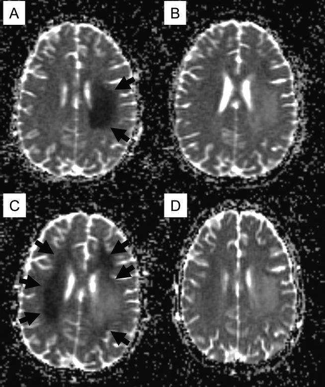
Fig. 1.
Diffusion-weighted imaging (apparent diffusion coefficient map) of the brain at the onset of subacute methotrexate (MTX) neurotoxicity (A), resolution of the initial episode (B), recurrent neurotoxicity after rechallenge with intrathecal MTX (C) and resolution of recurrent episode (D). Areas of restricted diffusion are shown in arrows.

