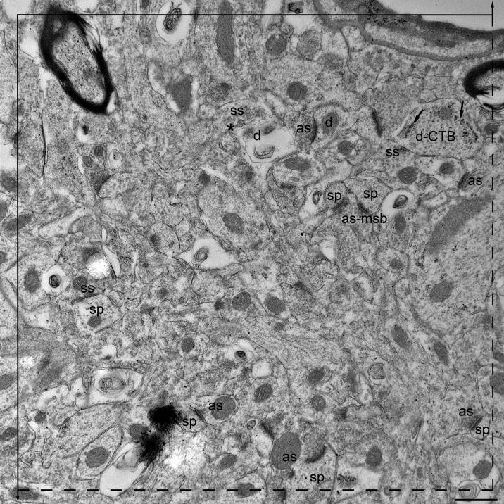
Figure 3A-B.
A pair of electron micrographs, taken from serial sections through the mPFC, in which synapses within the unbiased counting frame are labeled. Synaptic contacts are labeled according to their membrane specialization (as, asymmetric; ss, symmetric) and their postsynaptic targets (sp, spine; d, dendrite). Asterisks (*) mark the tops of synapses, i.e., those found in the reference (upper panel) but not the look up (lower panel) section. Multisynaptic boutons (msb) and CTB (marked with arrows) in the postsynaptic target are both labeled. Each micrograph was used as both the reference (3A) and look up (3B) section. Scale bar, 500 nm.


