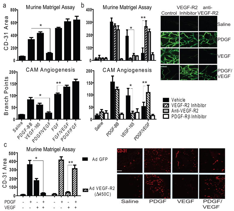
Figure 1. VEGF Inhibits PDGF-mediated Angiogenesis through VEGF-R2.
a, Upper: vessel growth into Matrigel as measured by CD-31+ area, *P<0.02. Bottom: branch points quantified on filter paper impregnated with saline or growth factors placed on embryonic day 10 Chick CAMs; *P<0.02, **p<0.05, results from one-way ANOVA. Further images are displayed in Supplementary Fig. 1d. b, Upper: vessel growth into Matrigel as in a, with the application of inhibitors; *P<0.001, **P<0.001. Bottom: branch points quantified on filter paper impregnated with saline or growth factor as in a, with the application of inhibitors; *P<0.02, **P<0.05 *results from two-way ANOVA. Right: confocal microscopy of Matrigel from mice injected intravenously with fluorescent Griffonia lectin; Scale bar, 100 μm c, CD-31+ area quantified by image analysis of Matrigel (right panel) impregnated with saline or growth factor implanted into mice infected with adenovirus expressing GFP only or GFP and truncated VEGF-R2 (Δ450C); Scale bar, 200 μm; *P<0.001, **P<0.001, results from one-way ANOVA. All error bars ± S.D; n>5 per group in all panels.

