
Figure 1.
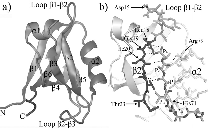
The structure of PDZ2 domain. (a) The overall structure of PDZ2 domain (PDB ID 3PDZ). Image was made with VMD 38. (b) An enlarged view of the ligand peptide and the binding pocket (PDB ID 1D5G). Helix α2, strand β2 and loop β1–β2 are colored yellow, blue and green, respectively. Hydrogen bonds between ligand peptide and PDZ2 domain are shown as dash lines. Images was made by Pymol 39.