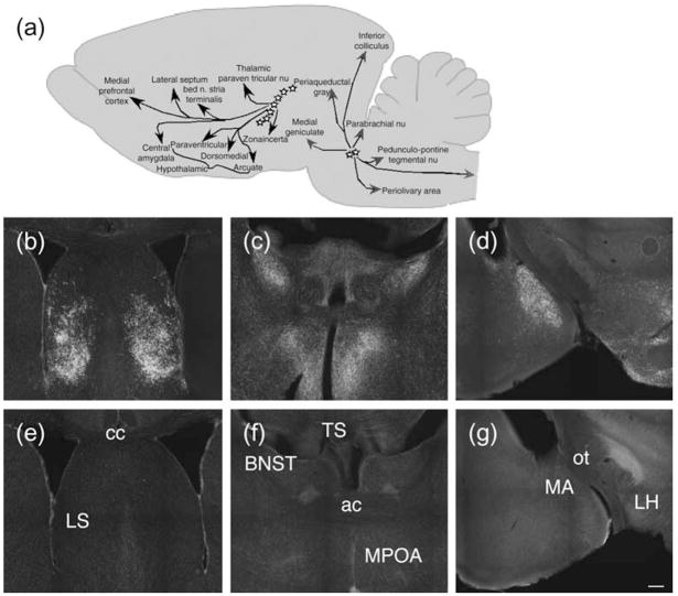
Figure 1. Absence of TIP39-like immunoreactivity in mutant mice.
(a) Cartoon illustrating location of TIP39 neurons and their major projections. (b–g) Antibody labeling in WT and TIP39-KO. Sections that include the lateral septum (b, e), bed nucleus of the stria terminalis and medial preoptic area (c, f), and parts of the amygdala (d, g) from WT (b–d) and age-matched TIP39-KO (e–g) were labeled with antibodies to TIP39. Note the absence of TIP39 labeling in sections from TIP39-KO (bottom row). LS, lateral septum; BNST, bed nucleus of the stria terminalis; TS, triangular septal nucleus; MPOA, medial preoptic nucleus; MA, medial amygdala; LH, lateral hypothalamus; cc, corpus callosum; ac, anterior commisure; ot, optic tract. Scale bar = 200 μm for all sections.

