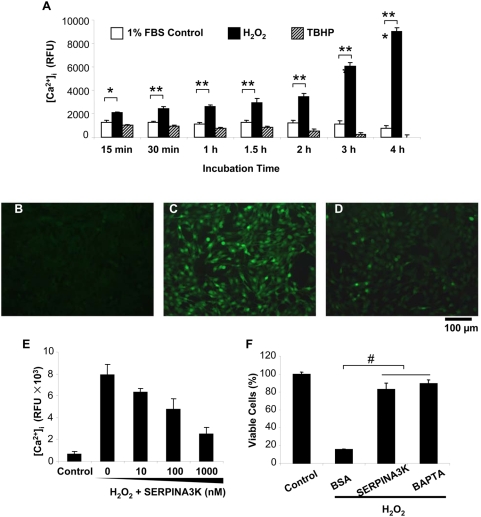
Figure 4. SERPINA3K blocks intracellular calcium overload induced by H2O2.
(A) Elevated intracellular [Ca2+] in cells exposed to H2O2. The Müller-derived rMC-1 cells were treated with 400 µM H2O2 for various durations as indicated. TBHP (20 µM) was used as control. Intracellular [Ca2+] was measured by the fluorescence of the probe Fluo-4/AM (mean±SEM, n = 8). (B–E) The cells were pre-treated with 1 µM SERPINA3K or BSA for 1 h followed by the H2O2 exposure. BSA was added to bring the total protein concentration to the same in each well. Representative fluorescence images were captured under a fluorescent microscope from untreated cells (B), cells pre-treated with 1 µM BSA (C) and with 1 µM SERPINA3K (D) followed by a 3-h exposure to H2O2. (E) [Ca2+] in cells pre-treated with various concentrations of SERPINA3K prior to the exposure to H2O2 (mean±SEM, n = 6). (F) The protective effects of 1 µM SERPINA3K and 10 µM BAPTA/AM (a calcium chelator) were quantified using the MTT assay (mean±SEM, n = 3). * P<0.05, ** P<0.01, *** P<0.001, the cells treated by H2O2 versus the control cells. # P<0.01, the SERPINA3K or BAPTA-treated cells versus the BSA-treated cells. Scale bar, 100 µm.

