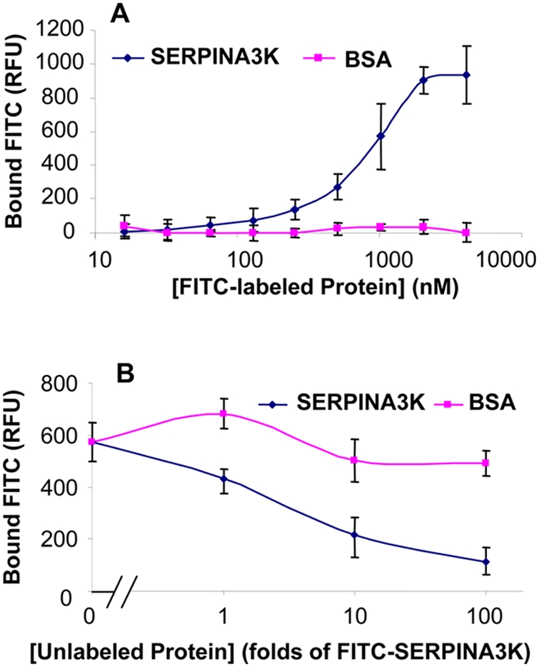
Figure 7. SERPINA3K binds to Müller-derived rMC-1 cells.

(A) The rMC-1 cells were incubated with increasing concentrations of FITC-SERPINA3K or FITC-BSA for 1 h. (B) The rMC-1 cells were incubated with 400 nM FITC-SERPINA3K in the presence of different concentrations of unlabeled SERPINA3K or BSA for 1 h. Then the cells were washed three times with PBS. The fluorescence in the cells was measured by a fluorometer using 485/530 nm filter (mean±SEM, n = 8).
