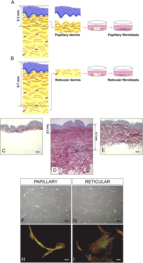
Figure 1. Experimental definition of Fp and Fr cells.
(A) and (B), principle of papillary (Fp) and reticular (Fr) fibroblast isolation. (C) Histological preparation of a mammary skin sample after dermatome cutting at a depth of 0.3 mm to obtain the superficial dermis with the epidermis. (D) Full thickness histology of the same skin sample. (E) Histological preparation of the skin sample after dermatome cutting at a depth of 0.7 mm to reach the deep dermis. Scale bars = 50 µm. (F) and (G) Photographs of typical cultures of Fp and Fr in light microscopy. Scale bars = 10 µm. (H) and (I) Fluorescence photographs of Fp and Fr after actin and vinculin immuno-staining. Scale bars = 2.5 µm.

