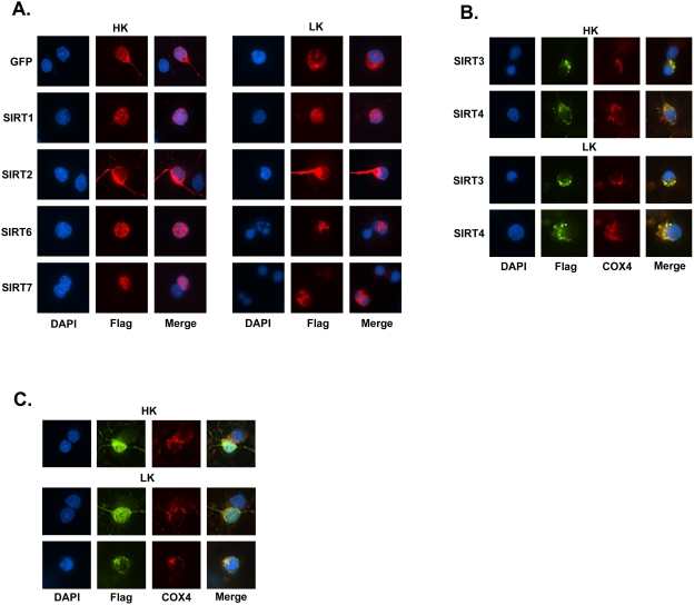
Figure 2. Subcellular localization pattern of SIRT 1–7 in neurons.
Expression plasmids encoding Flag-tagged forms of SIRT 1–7 were transfected into CGNs and the cultures were switched to HK or LK medium. The localization of each sirtuin was visualized by immunocytochemistry using a Flag antibody. DAPI staining was used to label the nucleus. In some cases, Cox4 antibody was used to visualize mitochondria. Images were obtained with a Nikon Eclipse 80i using a 60× objective. (A) Subcellular localization of SIRT1, SIRT2, SIRT6, and SIRT7 in HK and LK-treated neuronal cultures. Non-apoptotic cells were used for SIRT2 and SIRT6 in HK to show localization. (B) Mitochondrial localization of SIRT3 and SIRT4. The punctate pattern of SIRT3 and SIRT4 correspond to mitochondrial localization as evidenced by the overlap with Cox4 immunostaining. (C) Subcellular localization of SIRT5 in CGNs. In HK treatment SIRT5 predominately localizes to the cytoplasm, mitochondria, and nucleus (HK panel) while in LK SIRT5 can localize to the cytoplasm, mitochondria, and nucleus (upper LK panel) or to mitochondria (lower LK panel). Mitochondrial localization of SIRT5 is associated with apoptosis.

