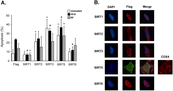
Figure 5. Forced sirtuin expression in the HT-22 cells.
(A) Effect of sirtuin expression on HT-22 cell survival. Sirtuin expression levels were enhanced in HT-22 neuroblastoma cells via transfection with mammalian expression plasmids. HT-22 cells were treated 24 hours following transfection with normal growth medium (untreated), 1 mM HCA or Serum-free (SF) medium for 24 hours. The viability of transfected cells was determined using a TUNEL assay. The mean of 5 experiments were taken (* indicates p<.05 compared with Flag untreated sample; # indicates p<.05 compared with Flag HCA sample; ˆ indicates p<.05 compared with Flag SF sample). (B) Subcellular localization of exogenous SIRT1, SIRT2, SIRT3, SIRT5 and SIRT6 in HT-22 cells. HT-22 cells were transfected with SIRT-Flag constructs and immunocytochemistry performed using a Flag antibody. DAPI staining was used to visualize nuclei/chromatin. SIRT5 shows colocalization with the mitochondrial protein Cox4.

